Preventive thermal examinations into body functioning, safe and pain-free for all women.
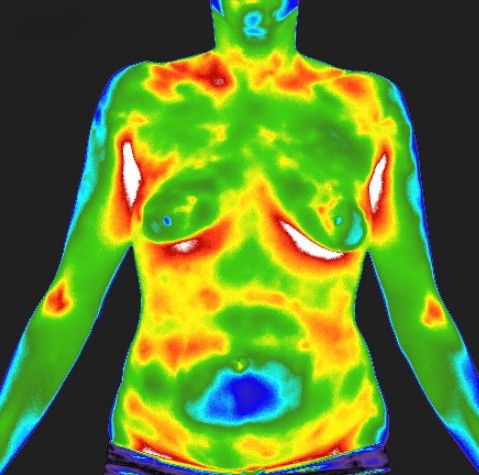
Medical thermography uses infrared technology to visualize and measure heat patterns on the body's surface. These patterns reflect inflammation, circulation, nerve function, and other physiological processes — offering a dynamic, non-invasive assessment that complements traditional diagnostic methods.
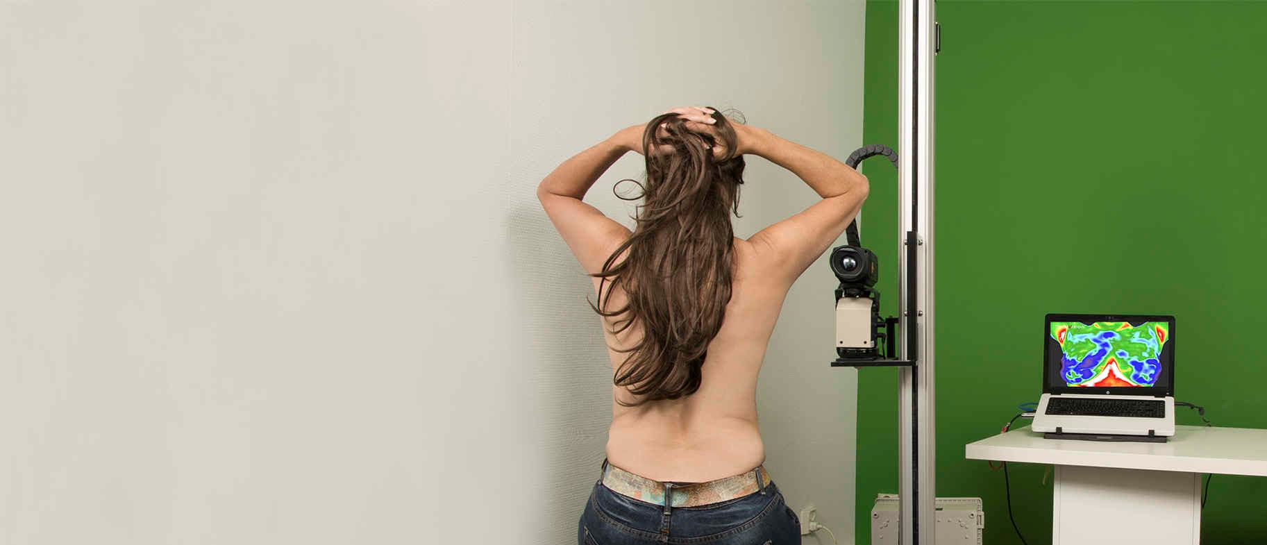
A consultation lasts between 15 and 60 minutes depending on the examination type. The procedure is completely painless, uses no radiation, and is suitable for women and men of all ages — including women with dense breast tissue, implants and women under 50 years.
Medical thermography is focused on physiology. It helps us observe functional changes that may be developing before structural changes are visible or diagnosable. Thermography is helping us pay attention to what is happening underground, at the level of the roots and the surrounding environment.
This is important because physiological stress and inflammation often precede structural disease. Thermography does not diagnose disease, but it can provide insight into early physiologic patterns that may suggest the need for closer monitoring or further investigation.
Let's use this analogy of a tree: a tree has roughly as much structure below the ground as it does above it. In many cases, conventional medicine identifies disease once it has already become structurally apparent — like changes seen in the trunk, branches, or leaves — because they are visible above ground.
A simple way to understand the difference is this: thermography looks at function, ultrasound looks at structure.
Medical thermography uses an infrared camera to visualize the heat patterns the body radiates through the skin. This creates a map of physiological activity. Think of differences in blood flow, temperature, asymmetry, and patterns that may be related to stress or recovery processes.
Ultrasound uses sound waves to visualize tissue structures. With ABUS ultrasound, systematic 3D images of both breasts are created. These images are subsequently reviewed by a radiologist. As a result, ultrasound has a very different role from thermography.
Therefore, the question is not: which examination is better? The better question is: what information do you want to obtain at this moment?
A safe, radiation-free examination method suitable for women from age 23. No compression, no discomfort.
The skin surface reflects internal metabolism and physiological changes. Thermography captures this without touching or penetrating the body.
Physicians assess signs of significant asymmetry indicating inflammation, blood perfusion, lymphatic congestion, and hormonal imbalance.
Gain deeper physiological insights beyond anatomical imaging. Monitor treatment effectiveness through thermal pattern changes.
Full-body exams offer comprehensive health monitoring, as the skin responds to physiological changes throughout the body's interconnected systems.
Thermography is an adjunct to other clinical diagnostic procedures. It provides complementary physiological information alongside other examination methods.
Thermography examinations are available through our affiliated practices.
The Green Precautionary Institute explicitly focuses on education, prevention, and scientific support. Our examinations are strictly preventative and intended for educational purposes only. We do not perform breast cancer screening, nor do we make medical diagnoses.